
Reference levels for diagnosis in nuclear medicine and hybrid imaging. Review and update
Article Sidebar
Main Article Content
Abstract
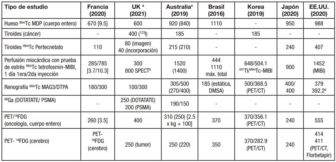
The diagnostic image must be carried out with the minimum necessary exposure of the patient that allows the objective of the diagnosis to be satisfactorily achieved. For this reason, diagnostic reference levels emerge and are established as dynamic tools to help optimize radiation protection, contribute to the standardization of practices and strengthen culture of safety, without compromising the clinical purpose of each examination or process. The objective of this work is to provide an updated overview of the establishment and use of these levels for nuclear medicine and hybrid imaging. It is identified that to establish and use them properly, trained personnel and coordination and collaboration activities are required among multiple actors, including medical services, health authorities, professional organizations and regulatory bodies. The accelerated development of technology generally exceeds the change in regulations, so these levels must be updated periodically, in order to fulfil their role as a guide and spur for optimization. The worldwide expansion of hybrid technologies and their growing use are a phenomenon of the last decade, so the establishment of these levels for such technologies has not been consolidated, although countries such as the United Kingdom and Australia show solid steps in this address. Research has been carried out with phantoms and directly with patients, the latter with a more useful contribution of information. The installation of hybrid equipment in Cuba demands this study, hence its importance.
Article Details

This work is licensed under a Creative Commons Attribution-NonCommercial 4.0 International License.
Aquellos autores/as que tengan publicaciones con esta revista, aceptan los términos siguientes:
- Los autores/as conservarán sus derechos de autor y garantizarán a la revista el derecho de primera publicación de su obra, el cuál estará simultáneamente sujeto a la Licencia Creative Commons Attribution-NonCommercial 4.0 International (CC BY-NC 4.0) que permite a terceros compartir la obra siempre que se indique su autor y su primera publicación esta revista. Bajo esta licencia el autor será libre de:
- Compartir — copiar y redistribuir el material en cualquier medio o formato
- Adaptar — remezclar, transformar y crear a partir del material
- El licenciador no puede revocar estas libertades mientras cumpla con los términos de la licencia
Bajo las siguientes condiciones:
- Reconocimiento — Debe reconocer adecuadamente la autoría, proporcionar un enlace a la licencia e indicar si se han realizado cambios. Puede hacerlo de cualquier manera razonable, pero no de una manera que sugiera que tiene el apoyo del licenciador o lo recibe por el uso que hace.
- NoComercial — No puede utilizar el material para una finalidad comercial.
- No hay restricciones adicionales — No puede aplicar términos legales o medidas tecnológicas que legalmente restrinjan realizar aquello que la licencia permite.
- Los autores/as podrán adoptar otros acuerdos de licencia no exclusiva de distribución de la versión de la obra publicada (p. ej.: depositarla en un archivo telemático institucional o publicarla en un volumen monográfico) siempre que se indique la publicación inicial en esta revista.
- Se permite y recomienda a los autores/as difundir su obra a través de Internet (p. ej.: en archivos telemáticos institucionales o en su página web) antes y durante el proceso de envío, lo cual puede producir intercambios interesantes y aumentar las citas de la obra publicada. (Véase El efecto del acceso abierto).
La Revista Nucleus solo aceptará contribuciones que no hayan sido previamente publicados y/o procesados, por otra publicación. Cualquier violación ese sentido será considerada una falta grave por parte del autor principal lo cual será objeto valoración por parte del Consejo Editorial, el cual dictaminará al respecto.
References
[2]. VITOLONI AC, LANCIONI F, MELADO G. Aporte de las imágenes híbridas SPECT/CT para el diagnóstico diferencial de lesiones óseas malignas y benignas en pacientes con cáncer de mama. Nuestra experiencia. Revista Argentina de Masteología. 2020; 39(41): 34-46.
[3]. KWON HW, KIM JP, LEE HJ, et. al. Radiation dose from whole-body F-18 fluorodeoxyglucose positron emission tomography/computed tomography: nationwide survey in Korea. J Korean Med Sci. 2016; 31Suppl 1(Suppl 1): S69-74. [consulta 10/07/2021]. Disponible en: http://dx.doi.org/10.3346/jkms.2016.31.S1.S69.
[4]. DAMILAKIS J, VASSILEVA J. The growing potential of diagnostic reference levels as a dynamic tool for dose optimization. Phys Med. 2021; 84:285-287. [consulta 10/07/2021]. Disponible en: https://doi.org/10.1016/j.ejmp.2021.03.018.
[5]. BERTOLINI V, PALMIERI A, BASSI MC, et. al. CT protocol optimisation in PET/CT: a systematic review. EJNMMI Phys. 2020; 7(1):17. [consulta 10/07/2021]. Disponible en: https://doi.org/10.1186/s40658-020-00287-x.
[6]. PAULO G, DAMILAKIS J, TSAPAKI V, et. al. Diagnostic reference levels based on clinical indications in computed tomography: a literature review. Insights Imaging. 2020; 11(1): 96. [consulta 10/07/2021]. Disponible en: https://doi.org/10.1186/s13244-020-00899-y.
[7]. ALKHYBARI EM, MCENTEE MF, BRENNAN PC, et. al. Determining and updating PET/CT and SPECT/CT diagnostic reference levels: a systematic review. University of Salford, United Kingdom, 2018. [consulta 10/07/2021]. Disponible en: http://usir.salford.ac.uk/id/eprint/47455/.
[8]. ALHAILIY AB, BRENNAN PC, MCENTEE MF, et. al. Diagnostic reference levels in cardiac Computed Tomography angiography: a systematic review. Radiat Protect Dosimetry. 2018; 178(1): 63-72. doi: 10.1093/rpd/ncx075.
[9]. UBEDA DE LA CARIDAD C, VAÑÓ E, RUIZ CRUCES R, et. al. Niveles de referencia para diagnóstico: una herramienta efectiva para la protección radiológica de pacientes. Rev Chil Radiol. 2019; 25(1): 19-25.
[10]. GIMELLI A, AIMO A. The relativity of reference values for myocardial perfusion imaging. JACC: Cardiovascular Imaging. 2021; 14(3): 666-668. [consulta 20/08/2021]. Disponible en: https://doi.org/10.1016/j.jcmg.2020.06.028.
[11]. MONTANGIE L, SANZ V, ILLANES L. Imágenes en medicina nuclear: verificación de su validez en la práctica cotidiana. Universidad Nacional de La Plata, Facultad de Ciencias Exactas, 2019.
[12]. SANZ V, MEDINA M, PALAU A, NAMÍAS M. Establecimiento de niveles de referencia institucionales para procedimientos de medicina nuclear. XXI Congreso Argentino de Medicina Nuclear. Buenos Aires, Argentina. 2018.
[13]. LOZAIN IG. Implementación de un sistema de gestión de dosis en estudios de tomografía computada [tesis de Maestría en Física Médica]. Argentina: Instituto Balseiro, 2018.
[14]. MARTÍNEZ GÓMEZ J, PÉREZ DÍAZ M, RUIZ GONZÁLEZ Y, FALCÓN RUIZ A. Análisis de calidad de imagen en PET/CT. Revista Cubana de Ciencias Informáticas 2017; 11(1): 29-40. [consulta 10/07/2021]. Disponible en: http://rcci.uci.cu.
[15]. Ministerio de Ciencia, Tecnología y Medio Ambiente y Ministerio de Salud Pública CITMA/MINSAP). Normas básicas de seguridad radiológica. Resolución conjunta. La Habana, 2001.
[16]. VAÑÓ E, MILLER DL, MARTIN CJ, et al. ICRP Publication 135: Diagnostic reference levels in medical imaging. Ann ICRP. 2017; 46(1): 1-144.
[17]. Organismo Internacional de Energía Atómica (OIEA). Protección radiológica y seguridad de las fuentes de radiación: Normas básicas internacionales de seguridad. Requisitos de seguridad generales. Parte 3. Viena: OIEA, 2016.
[18]. VASSILEVA J. DRLs in diagnostic NM and hybrid imaging. Patient exposure monitoring. Regional therapy center on radiation protection in diagnostic and therapeutic nuclear medicine regional training course. Valeta, Malta. 2019.
[19]. International Atomic Energy Agency, International Labour Office, Pan American Health Organization and World Health Organization (IAEA/ILO/PAHO/WHO). Radiation protection and safety in medical uses of ionizing radiation. Safety Guide, SSG-46. Vienna: IAEA, 2018.
[20]. VASSILEVA J. Joint ICTP-IAEA Workshop on establishment and utilization of diagnostic reference levels in medical imaging. Nov 2019. Vienna, Austria. 2019 [consulta 10/07/2021]. Disponible en: https://www.iaea.org/newscenter/news/more-resourcesare-needed-for-developing-and-using-diagnostic-referencelevels.
[21]. VASSILEVA J. Diagnostic reference levels (DRLs): the concept and use. 3th International Conference on Dosimetry and its Applications. ICDA 3. 27-31 May 2019. Lisbon, Portugal. [consulta 10/07/2021]. Disponible en: www.slideshare.net›diagnostic-reference-levels-drls-the-concept-and-use.
[22]. Agency International Atomic Energy (IAEA). Webinar series on the establishment and utilization of diagnostic reference levels in medical imaging in Europe 2021 [consulta 25/08/2021]. Disponible en: https://www.iaea.org/resources/webinar/webinar-series-on-the-establishmentand-utilization-of-diagnostic-referencelevels-in-medical-imaging-in-europe.
[23]. Agency International Atomic Energy (IAEA). IAEA Learning management system. Diagnostic reference levels in medical imaging [website]. 2021 [consulta 12/09/2021]. Disponible en: https://elearning.iaea.org/m2/course/index.php?categoryid=148.
[24]. Agency International Atomic Energy (IAEA). Radiation protection of patients in nuclear medicine: diagnostic reference levels and accuracy of activity meters. [consulta 12/07/2022]. Disponible en: https://www.iaea.org/resources/webinar/radiation-protection-of-patients-in-nuclear-medicine-diagnostic-reference-levels-and-accuracy-of-activity-meters.
[25]. VASSILEVA J. Diagnostic reference levels in diagnostic nuclear medicine. Patient exposure monitoring. Virtual Joint ICTP-IAEA Workshop on Radiation Protection in Diagnostic and Therapeutic Nuclear Medicine (SMR 3633). 11- 15 Oct 2021. Italy.
[26]. MAHESH M, MORIN RL. The DICOM radiation dose structured report: what it is and what it is not. J Am Coll Radiol. 2015; 12(7): 712-713. [consulta 10/07/2021]. Disponible en: http://dx.doi.org/10.1016/j.jacr.2015.04.002:712-3.
[27]. Sociedad Española de Protección Radiológica. Requisitos básicos para los sistemas de registro y gestión de dosis en pacientes sometidos a exploraciones de diagnóstico por imagen. 2020 [consulta 10/07/2021]. Disponible en: https://www.sepr.es/profesionales/descargables/download/29-control-de-calidad-en-radiodiagnostico/4380-requisitos-basicos-para-los-sistemas-de-registro-y-gestion-de-dosis-en-pacientes-sometidos-a-exploraciones-de-diagnostico-por-imagen.
[28]. YASEEN A-BB, RADZI YMD, ALMOHIY HM, et. al. Strategies to improve CT dose optimization for hybrid PET/CT imaging. Open Journal of Medical Imaging. 2021 [consulta 20/08/2021]; 11:48-57. Disponible en: https://www.scirp.org/journal/ojmi.
[29]. GARDNER M, KATSIDZIRA NM, ROSS E, LARKIN EA. Patient dosimetry audit for establishing local diagnostic reference levels for nuclear medicine CT. Br J Radiol. 2017 [consulta 10/07/2021]; 90: 20160850. Disponible en: https://doi.org/10.1259/bjr.20160850.
[30]. American Association of Physicists in Medicine (AAPM). Size-specific dose estimate (SSDE) for heat CT. AAPM REPORT NO. 293. July, 2019 [consulta 10/07/2021]. Disponible en: https://www.aapm.org/pubs/reports/?s=204&submit=submit.
[31]. HU X, GOU J, LIN W, ZOU C, LI W. Size-specific dose estimates of adult, chest computed tomography examinations: comparison of Chinese and updated 2017 American College of Radiology Diagnostic Reference Levels based on the water equivalent diameter. PLoS ONE. 2021; 16(9): e0257294 [consulta 10/07/2021]. Disponible en: https://doi.org/10.1371/journal.pone.0257294.
[32]. RAJARAMAN V, PONNUSAMY M, HALANAIK D. Size specific dose estimate (SSDE) for estimating patient dose from CT used in myocardial perfusion SPECT/CT. Asia Ocean J Nucl Med Biol. 2020; 8(1): 58-63. doi: 10.22038/aojnmb.2019.40863.1276.
[33]. IBALL GR, BEBBINGTON NA, BURNISTOND M, et. al. A national survey of computed tomography doses in hybrid PET-CT and SPECT-CT examinations in the UK. Nucl Med Comm. 2017; 38(6): 459-70. doi: 10.1097/MNM.0000000000000672.
[34]. ALKHYBARI EM, MCENTEE MF, BRENNAN PC, et. al. Diagnostic reference levels for 18F-FDG whole body PET/CT procedures: results from a survey of 12 centres in Australia and New Zealand. Med Imaging Radiat Oncol. 2019; 63(3): 291-299.
[35]. Agency Australian Goverment. Australian radiation protection and nuclear safety. Australian diagnostic reference levels (DRLs) for nuclear medicine. Technical report No. 180. 2019 [consulta 10/07/2021]. Disponible en: https://www.arpansa.gov.au/research-and-expertise/surveys/national-diagnostic-reference-level-service/nm/in-more-detail.
[36]. ROCH P, CELIER D, DESSAUD C, ETARD C. Patient exposure from nuclear medicine in France: National follow-up and influence of the technology through diagnostic reference levels data analysis. Radiat Protect Dosim. 2017; 1(179): 87-94. doi: 10.1093/rpd/ncx213.
[37]. Institutee Radioprotection et de Sureté Nucléare (IRSN). Analysis of data for updating diagnostic reference levels in radiology and nuclear medicine. 2016-2018, Report. 2020.
[38]. DEBELJUH DD, JURKOVIC S, PRIBANIC I, et. al. National survey to set diagnostic reference levels in nuclear medicine single photon emission imaging in Croatia. Phys Med. 2020; 78: 109-116. [consulta 10/07/2021]. Disponible en: https://doi.org/10.1016/j.ejmp.2020.09.005.
[39]. NIKSIRAT F, MONFARED AS, DEEVBAND MR, et. al. Estimating the population dose from nuclear medicine examinations towards establishing diagnostic reference levels. Indian J Nucl Med. 2016; 31(1): 31-5. doi: 10.4103/0972-3919.172353
[40]. ALI WM, ELAWAD RM, IBRAHIM MAA. Establishment of dose reference levels for nuclear medicine in Sudan. Open Journal of Radiology. 2016; 6(4): 258-263 [consulta 10/07/2021]. Disponible en: http://dx.doi.org/10.4236/ojrad.2016.64034.
[41]. SALAMA DH, VASSILEVA J, MAHDALY G, et. al. Establishing national diagnostic reference levels (DRLs) for computed tomography in Egypt. Phys Med. 2017; 39: 16-24. [consulta 10/07/2021]. Disponible en: http://dx.doi.org/10.1016/j.ejmp.2017.05.050.
[42]. DE PONTI E. Exposure monitoring and DRLs in diagnostic nuclear medicine and hybrid imaging: quantities, procedures, methods. Italian experience with DRLs for nuclear medicine. 18-22 November 2019. Trieste, Italy.
[43]. ABE K, HOSONO M, IGARASHI T, et, al. The 2020 national diagnostic reference levels for nuclear medicine in Japan. Ann Nucl Med. 2020; 34(11): 799-806. [consulta 10/07/2021]. Disponible en: https://doi.org/10.1007/s12149-020-01512-4.
[44]. SAGARA H, INOUE K, YAKU H, et. al. Optimization of injection dose in 18F-FDG PET/CT based on the 2020 national diagnostic reference levels for nuclear medicine in Japan. Ann Nucl Med. 2021; 35(11):1177-1186. doi: 10.1007/s12149-021-01656-x.
[45]. LIMA TVM, GNESIN S, RYCKX N, et. al. Review Swiss survey on hybrid imaging CTs doses in Nuclear Medicine and proposed national dose reference levels. Z Med Phys. 2018; 28(4): 265-275. [consulta 10/07/2021]. Disponible en: https://doi.org/10.1016/j.zemedi.2018.01.005.
[46]. SONG HC, NA MH, KIM J, et. al. Diagnostic Reference levels for adult nuclear medicine imaging established from the national survey in Korea. Nucl Med Mol Imag. 2019; 53(1): 64-70. [consulta 10/07/2021]. Disponible en: https://doi.org/10.1007/s13139-019-00585-y.
[47]. American College of Radiology (ACR)/American Association of Physicists in Medicine (AAPM)/American College of Nuclear Medicine (ACNM)/Society of Nuclear Medicine and Molecular Imaging (SNMMI). ACR-AAPM-ACNM-SNMMI practice parameter for reference levels and achievable administered activity for nuclear medicine and molecular imaging. ACR-AAPM-ACNM-SNMMI, 2020.
[48]. NARAWONG T, SINGUSAHA O, SISAI S, et. al. The survey of local diagnostic reference levels (DRLs) for administered activity in diagnostic nuclear medicine at Rajavithi Hospital. Thai J Rad Tech. 2021; 46(1): 1-7.
[49]. STEPHENS T. OP-610. Investigation into the impact manufacturers have upon syringe residues in different volumes of radiopharmaceuticals. European Journal of Nuclear Medicine and Molecular Imaging. 2020; 47(1): S308.
[50]. SHAHZAD A, BASHIR S, ANWAR A. Establishment of age-specific reference levels and achievable doses for children and adults undergoing nuclear medicine exams. Radioprotection. 2019; 54(3):187-194 [consulta 10/07/2021]. Disponible en: https://doi.org/10.1051/radiopro/2019019.
[51]. SHAHZAD A, BASHIR S. Applications of diagnostic reference levels of standard doses in nuclear medicine. In: Nuclear Medicine Physics. London: IntechOpen; 2019. Available from: https://doi.org/10.5772/intechopen.87966.
[52]. FERREIRA GONÇALVES JM. Establishment of nuclear medicine diagnostic reference levels (DRL’s) in a large oncology hospital. [thesis Mestrado em Física Médica]. Universidade de Porto, 2019.
[53]. BECKER MD, BUTLER PF, SIAM M, et. al. US PET/CT and gamma camera diagnostic reference levels and achievable administered activities for noncardiac nuclear medicine studies. Radiology. 2019; 293(1): 203-211. [consulta 10/07/2021]. Disponible en: https://doi.org/10.1148/radiol.2019190623.
[54]. WILLEGAIGNON J, BRAGA LFFF, SAPIENZA MT, COURA-FILHO GB, ARDONA MAR, ALVES CER, et al. Diagnostic reference level: an important tool for reducing radiation doses in adult and pediatric nuclear medicine procedures in Brazil. Nuclear Medicine Communications. 2016; 37:525-33
[55]. RINSCHEID A, JANZEN T, ALIKHANI B, et. al. Radiation doses from low-dose CT scans in SPECT/CT and PET/CT examinations: a survey in Germany. Nuklearmedizin. 2022; 61(4): 294-300. doi: 10.1055/a-1759-3900.
[56]. VERFAILLIE G, D’ASSELER Y, BACHER K. European diagnostic reference levels for Computed Tomography applications in nuclear medicine: results from the MEDIRAD project. Eur J Nucl Med Mol Imag. 2020; 47(Suppl. 1): S79.
[57]. VERFAILLIE G, BACHER K. MEDIRAD. Deliverable 2.10 European DRLs for specific applications of CT in multi-modality systems. Project title: Implications of Medical Low Dose Radiation Exposure. Grant Agreement Number: 755523. 2020. doi: 10.1007/s00259-020-04988-4.
[58]. DENNIS JL, GEMMELL AJ, NICOL AJ. Optimization of the CT component of SPECT-CT and establishment of local CT diagnostic reference levels for clinical practice. Nucl Med Comm. 2018; 39(6): 493-499. doi: 10.1097/MNM.0000000000000831.
[59]. MANIVANNAN J, SHARMA R, GIRONELLA RM, PARTHIPUN A. Setting up local dose reference limits for orthopaedic bone SPECT-CT [website]. Trinity Medical Imaging, 2021. [consulta 15/10/2021]. Disponible en: https://www.google.com/url?sa=t&rct=j&q=&esrc=s&source=web&cd=&ved=2ahUKEwiRsZSrmvfzAhWASTABHdmyDdY4FBAWegQIChAB&url=https%3A%2F%2Fwww.nuclearmedicineblog.com%2Fwp-content%2Fuploads%2F2021%2F05%2FSetting-Up-Local-Dose-Reference-Limits-For-Orthopaedic-Bone-SPECT-CT-small.pdf&usg=AOvVaw0UXpqDv7nzxINaaF-kRLGz.
[60]. SAMARTZIS A, FOTEINA A, TZAMPAZIDOU E, et. al. Diagnostic reference levels of the combined whole-body PET/CT examinations in a large territory hospital of Greece. Congress ECR 2019. Poster Number: C-2578. [consulta 15/10/2021] 2019. Disponible en: https://epos.myesr.org/poster/esr/ecr2019/C-2578/Aims%20and%20objectives#poster.
[61]. DEIDDA D, THOMAS BA, FERREIRA K, et. al. Validation of SPECT-CT image reconstruction for the Mediso AnyScan SCP scanner in STIR. 2019 IEEE Nuclear Science Symposium and Medical Imaging Conference (NSS/MIC). [consulta 10/07/2021]. Disponible en: http://mrtdosimetry-empir.eu/.
[62]. POLI GL, COCA M, TORRES L, et. al. Developing and implementing an imaging optimization study in pediatric nuclear medicine: experience and recommendations from an IAEA Coordinated Research Project. J Nucl Med. 2021; 62(4): 570-576. doi: 10.2967/jnumed.120.244616.
[63]. TULIK M, TULIK P, KOWALSKA T, KOWALSKA T. On the optimization of bone SPECT/CT in terms of image quality and radiation dose. J Appl Clin Med Phys. 2020; 21(11):237-46. Doi: 10.1002/acm2.13069.
[64]. GROSSER OS, RUF J, KUPITZ D, et. al. Iterative CT reconstruction in abdominal low-dose CT used for hybrid SPECT-CT applications: effect on image quality, image noise, detectability, and reader’s confidence. Acta Radiologica Open. 2019; 8(6): 1-9. [consulta 10/07/2021]. Disponible en: http://dx.doi.org/10.1177/2058460119856266.
[65]. PRIETO E, GARCÍA-VELLOSO MJ, DÁMASO AQUERRETA J, et. al. Ultra-low dose whole-body CT for attenuation correction in a dual tracer PET/CT protocol for multiple myeloma. Physica Medica. 2021 [consulta 10/07/2021]; 84 1-9. Disponible en: https://doi.org/10.1016/j.ejmp.2021.03.019.
[66]. BEBBINGTON NA, JØRGENSEN T, DUPONT E, MICHEELSEN MA. Validation of CARE kV automated tube voltage selection for PET-CT: PET quantification and CT radiation dose reduction in phantoms. EJNMMI Physics. 2021 [consulta 10/07/2021]; 8(29):1-14. Disponible en: https://doi.org/10.1186/s40658-021-00373-8.
[67]. POLFLIET G. Optimization of CT radiation dose and image quality in hybrid nuclear medicine imaging [thesis of Master of Biomedical Sciences]. Ghent University, Germany, 2019.
[68]. SÖDERBERG M. Image quality optimisation and dose management in CT, SPECT/CT, and PET/CT. Thesis for the Degree of Doctor of Philosophy in Medical Science. Malmö, Sweden: Lund Universtiy; 2012. ISSN 1652-8220.
[69]. SHON IH, REECE C, HENNESSY T, HORSFIELD M, MCBRIDE B. Influence of X-ray computed tomography (CT) exposure and reconstruction parameters on positron emission tomography (PET) quantitation. EJNMMI Phys. 2020; 7(62): 2-16. [consulta 10/07/2021]. Disponible en: https://doi.org/10.1186/s40658-020-00331-w.
[70]. SOOKPENG S, MARTIN CJ, GENTLE DJ, LOPEZ-GONZALEZ MR. Relationships between patient sizes, dose and image noise under automatic tube current modulation systems. J Radiol Prot. 2014; 34: 103-23. doi: 10.1088/0952-4746/34/1/103.
[71]. European Commission. European study on clinical diagnostic reference levels for X-ray medical imaging: EUCLID. Radiation Protection No. 195. Luxembourg; 2021.
[72]. American Association of Physicists in Medicine (AAPM). The measurement, reporting, and management of radiation dose in CT. Report No. 96. AAPM, 2008.
[73]. American Association of Physicists in Medicine (AAPM). Performance evaluation of Computed Tomography systems. Report No. 233. AAPM, 2019.
[74]. AGUIRRE HURTADO CA. Dosis efectiva en pacientes adultos en tomografía computarizada [tesis de Maestría en Física Médica]. Pontificia Universidad Javeriana, Facultad de Ciencias, Departamento de Física Bogotá D.C, Colombia. Julio de 2021.
[75]. FABRI D, SOFFIA P, SANGUESA J, CASTILLO R. Establishing dose reference levels for CT in a Chilean hospital including technologist performance. Congress EuroSafe Imaging 2019. [consulta 10/07/2021]. Disponible en: https://epos.myesr.org/poster/eurosafe/eurosafeimaging2019/ESI-0101.
[76]. WANG KC, PATEL JB, VYAS B, et. al. Use of radiology procedure codes in health care: the need for standardization. RadioGraphics. 2017; 37(4):1099-1110. [consulta 20/08/2021]. Disponible en: https://doi.org/10.1148/rg.2017160188.
[77]. Administration of Radioactive Substances Advisory Committee (ARSAC). Notes for guidance on the clinical administration of radiopharmaceuticals and use of sealed radioactive sources. ARSAC, 2021 [consulta 20/08/2021]. Disponible en: http://www.gov.uk/arsac.
[78]. BANKS KP, GUNTHER RS, FARRELL MB, et al. U.S. Diagnostic reference levels and achievable administered activities for adult renal scintigraphy: an analysis of the Intersocietal Accreditation Committee Nuclear Laboratories. J Nucl Med Technol. 2021; 49(3): 246-249. [consulta 20/08/2021]. Disponible en: https://doi.org/10.2967/jnmt.120.261552.
[79]. American College of Radiology (ACR)/American Association of Physicists in Medicine (AAPM)/American College of Nuclear Medicine (ACNM)/Society of Nuclear Medicine and Molecular Imaging (SNMMI). Practice parameter for diagnostic reference levels and achievable doses in medical X-Ray imaging. ACR-AAPM-SPR, 2018 [consulta 20/08/2021]. Disponible en: https://www.acr.org/-/media/ACR/Files/Practice-Parameters/diag-ref-levels.pdf.
[80]. HIRSCHFELD C, DONDI M, PASCUAL T, et. al. Worldwide diagnostic reference levels for Single-Photon Emission Computed Tomography Myocardial Perfusion Imaging. JACC: Cardiovasc Imaging. 2020; 14(3): 657-665.
[81]. International Commission of Radiation Protection (ICRP). Radiation dose to patients from radiopharmaceuticals: a compendium of current information related to frequently used substances. ICRP Publication 128. Ann. ICRP 44(2S).
[82]. Red Latinoamericana de Protección Radiológica (LAPRAM). Webinar “Optimización en tomografía computada mediante el uso de DRL y la importancia de incorporar la cuantificación de la calidad de la imagen”. 21 mayo, 2019. Santiago de Chile. [consulta: 10/07/2021]. Disponible en: https://www.youtube.com/watch?v=tLNJYHxx9co.
[83]. VAÑÓ E, FRIJA G, LOOSE R, et. al. Dosimetric quantities and effective dose in medical imaging: a summary for medical doctors. Insights into imaging. 2021; 12: 99. [consulta: 20/08/2021]. Disponible en: https://doi.org/10.1186/s13244-021-01041-2.